X-Ray-
|
X-Rays have a great penetrating power, although they are invisible. They are used in investigating the structure of objects and to find the flaws in metals. This ray has the power to damage cells and tissues, as well as the genetic material inside of the cells.
X-Rays are produced when an electrons hit a metal surface. This happens in an X-Ray tube. X-Rays are also used in radiology to observe and create an image of the structure of bones. They are further used in radiotherapy, where X-Rays are fired at cancer cells in order to kill them or stop them from reproducing. X-rays have very small wavelengths, between 0.03 and 3 nanometers, so small that some x-rays are no bigger than a single atom of many elements. They have frequencies in the range 30 petahertz to 30 exahertz. People that work with x-rays, it is important that they use protective lead shields and monitor their exposure levels. The latter can be done so with a Personal Radiation Monitoring Device or a PMD. It is the responsibility of the employee to monitor their levels of exposure, to prevent the worker from possible harm. What stops x-rays? X-rays are absorbed by dense materials such as lead and uranium, due to the fact that they can penetrate only a short distance into the material. This is why lead shielding is used to protect people from excessive exposure to x-rays. History of X-rays X-rays were first observed and documented in 1895 by a German scientist by the name of Wilhelm Conrad Roentgen. He discovered that firing streams of x-rays through the arms and hands of people created detailed images of the bones inside. When you get an x-ray taken, an x-ray sensitive film is put on one side of your body, and x-rays are shot through you. Because our bones are dense and absorb more x-rays than skin does, the shadow of the bones are left on the x-ray film while the skin appears transparent, allowing us to easily view any damage or objects within the body. Ionising Because x-rays have so much energy due to their short wavelength, they can ionise atoms they target. It is likely, that it will do little or no harm, but when atoms in living cells are ionised, it can kill the cell or cause mutations. For this reason, exposure to x-rays should be controlled to keep the level of exposure in a safe range. |
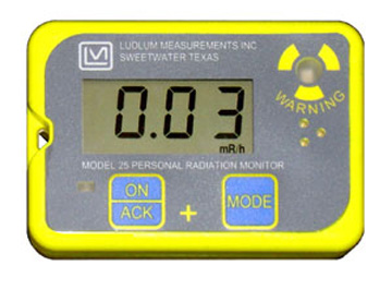
Personal Radiation Monitoring Device
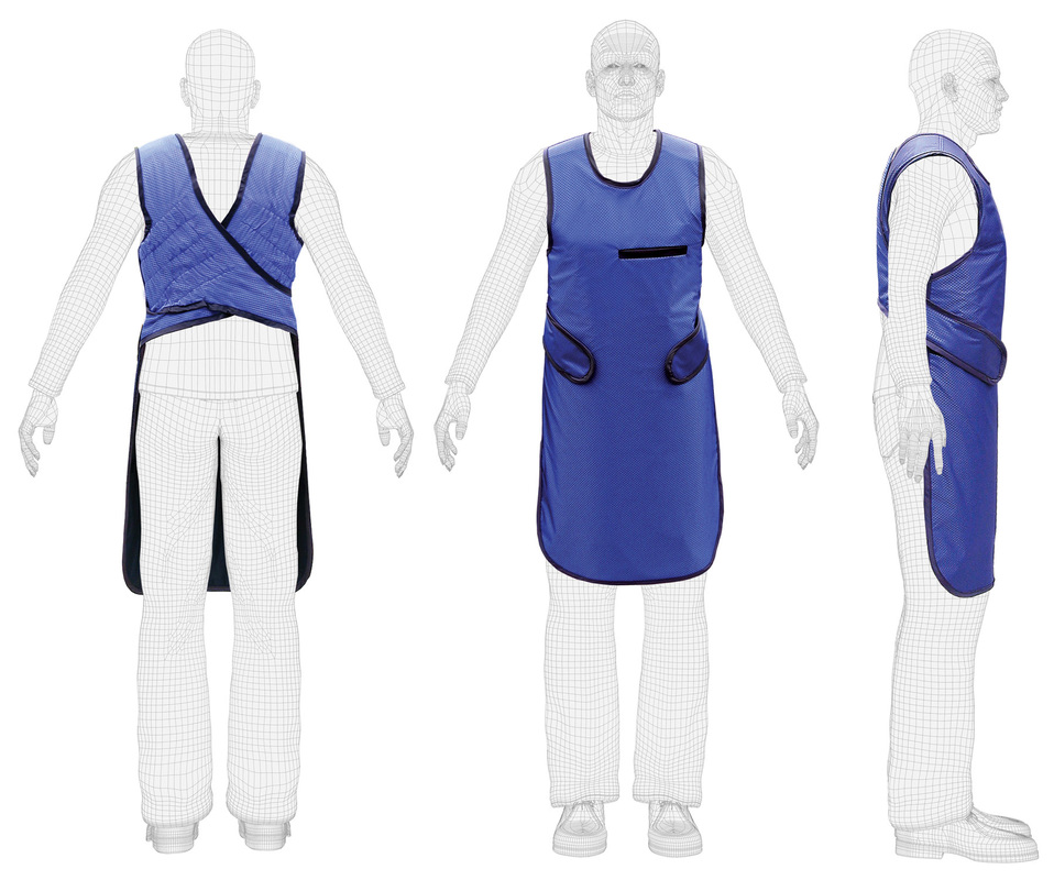
Lead shield.
|
|
In today's society we use:
Fluoroscopy A fluoroscopy unit uses x-rays and an Image Intensifier to produce ‘live’ 2D images, similar to that of a movie. Often, a contrasting agent (barium or iodine based) is used make different body parts more visible, for easier viewing. Esentially, a continuous X-ray beam is passed through the body part being examined. A fluoroscopy may be performed to evaluate specific areas of the body, including the bones, muscles, and joints, as well as solid organs, such as the heart, lung, or kidneys. This allows for fluoroscopy to be used as a diagnostic procedure, or it may be used in conjunction with other diagnostic or therapeutic procedures. Furthermore, this technology provides doctors with an easy method to view the internals of a patient who may be experiencing pain in parts of their body that woukd be otherwise extremely hard to reach. |
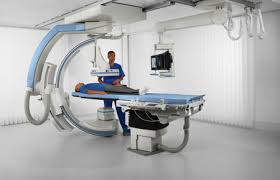
Fluoroscopy machine.
|
